Back to: Neurology
Dural Haematomas
Extradural Haematoma
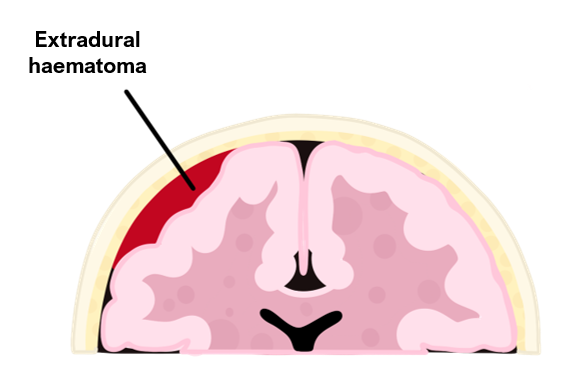
An extradural haematoma is a bleed into the “potential” space between the dura mater meningeal layer and the inner surface of the skull.
It usually occurs due to low impact trauma to the side of the head, resulting in laceration of the middle meningeal artery running deep to the pterion.
This causes the dura to peel away from the inner layer of the skull. As the bleed enlarges it compresses the brain and raises intracranial pressure causing symptoms.
Symptoms
Initial headache and loss of consciousness
Causes a lucid interval (hours to days) as the haematoma grows
This is followed by rapid deterioration with severe headache, decreasing GCS and seizures
Pressure symptoms – CN III palsy (ipsilateral pupil dilates), leg weakness, irregular breathing
Raised ICP causes Cushing’s triad – bradycardia, hypertension and deep breathing
In untreated, can lead to respiratory arrest (compression of medulla)
Key tests
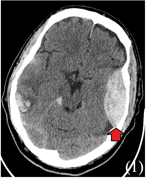
Non-contrast CT is first-line – shows biconvex, high-density area or “lemon” sign which does not cross suture lines
Management
Reversal of anticoagulation, if needed
Medication involves diuretics (to reduce ICP) and prophylactic antibiotics
Surgery – craniotomy at haematoma to relieve pressure and ligation of blood vessels
Subdural Haematoma
This is a collection of blood between the dura and arachnoid mater of the brain.
It is categorised as acute (< 3 days, associated with high impact trauma), subacute (3–21 days), or chronic (> 21 days, more common in elderly and alcoholics).
Can be due to high impact trauma to head, rupturing the bridging veins in the dural venous sinuses, but also secondary to metastases and rupture of aneurysms.
Risk factors
Elderly (brain atrophy makes bridging veins vulnerable)
Alcoholism
Anticoagulation
Symptoms
Chronic headache or new onset headache (if acute)
Fluctuating level of consciousness is a key symptom
Confusion and altered mental state (irritability)
Localising neurological signs occur late, e.g., unequal pupils, loss of smell/ hearing
Key tests
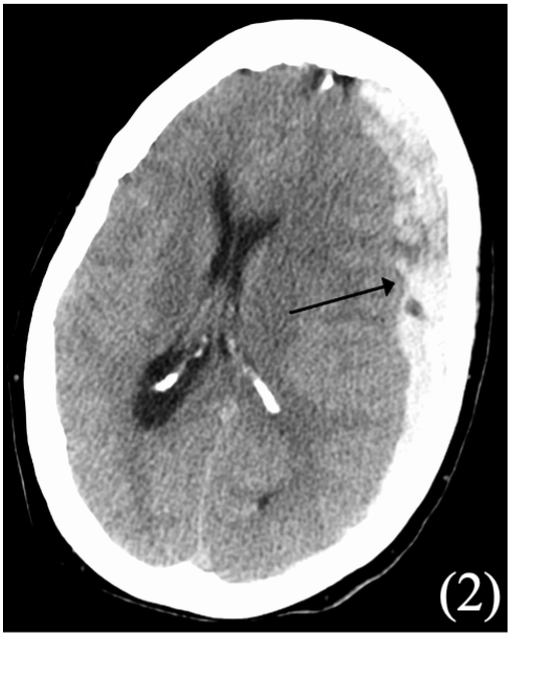
Non-contrast CT shows concave “banana” shape (does cross suture lines)
Management
Reverse clotting abnormalities
Antiepileptics may be started if causing seizures
If small, can be treated conservatively and monitored for resolution
If large or symptomatic, consider surgery (decompressive craniotomy)
NICE Guidelines for CT head - Adults
CT scan immediately if:
– GCS <13 or <15 2 hours post-injury
– Any seizure
– Any neurological deficit
– >1 episode of vomiting
– Any sign of skull fracture (hemotympanum, “panda” eyes, CSF lead from ear/nose, Battle’s sign)
CT within 8 hours if:
– Age >65
– History of bleeding/clotting disorder or taking warfarin
– Fall height >1m or 5 stairs
– >30 mins retrograde amnesia of events before injury
NICE Guidelines for CT head - Children
CT scan immediately if:
– Loss of consciousness > 5mins
– Amnesia > 5 mins
– >2 episodes of vomiting
– Post-traumatic seizure
– Any sign of skull fracture
– Any neurological deficit
– Fall > 3m
– GCS <14, or less than 15 if baby under 1 year
– If under 1, presence of bruise, wound >5cm on head
Sources
2. James Heilman, MD / CC BY-SA (https://creativecommons.org/licenses/by-sa/3.0)

