




















































Back to: Dermatology
Birth Marks
Children often present with a variety of birthmarks – these can be asymptomatic or develop into diseases. They can be categorised by their colour.
Blue birthmarks
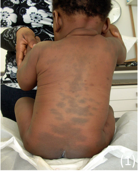
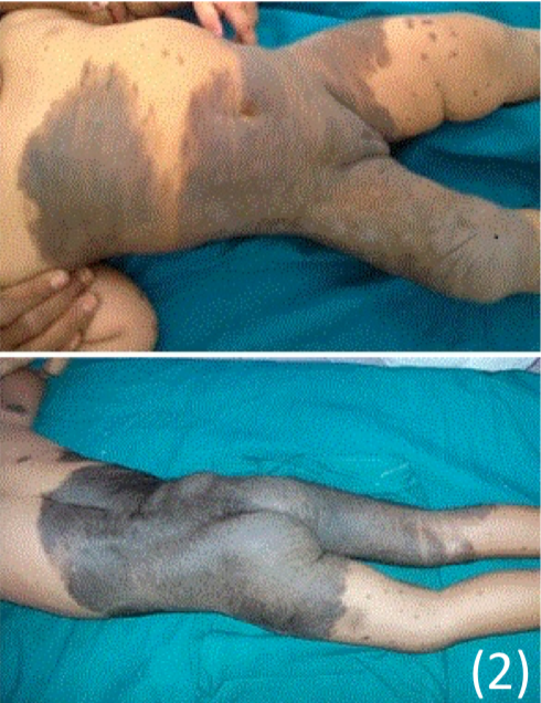
Mongolian Blue spots
These are congenital marks, which occur mainly in Asian/African children
– Colour occurs as dermal melanocytes become interrupted in their migration from neural crest to epidermis in utero. They are completely asymptomatic
Appearance – Blue-grey macules, usually lumbosacral
Brown birthmarks
Congenital Melanocytic naevi
These are congenital marks which occur due to congenital proliferation of melanocytes
Appearance
– Present as a brown plaque which can be hairy.
– Size ranges Small/medium/ Large/ giant
Giant congenital melanocytic naevi (GCMN), are associated with other conditions:
– Neurocutaneuous melanosis –> melanocyte proliferation into the CNS
– Melanoma
– Ulceration
The malignancy risk of GCMN is 15% in giant lesions, especially those which have “satellite” lesions
– 70% of the malignancy occurs before puberty, and can be extra-cutaneous (involving the CNS)
Red birthmarks
These types of birthmarks get their red colour as they involve the vascular system. They are split into two categories which are often confused:
Vascular tumours
This refers to a benign or malignant tumour (abnormal growth) of the cells forming the arteries, veins and capillaries. They are not clinically present at birth and have a period of rapid growth.
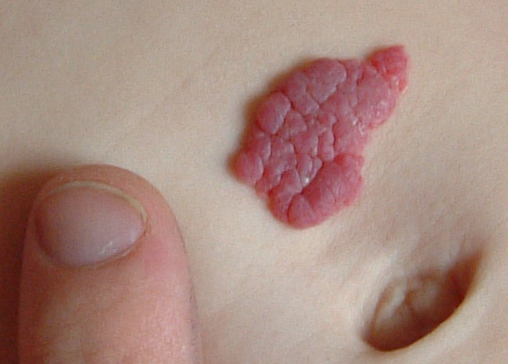
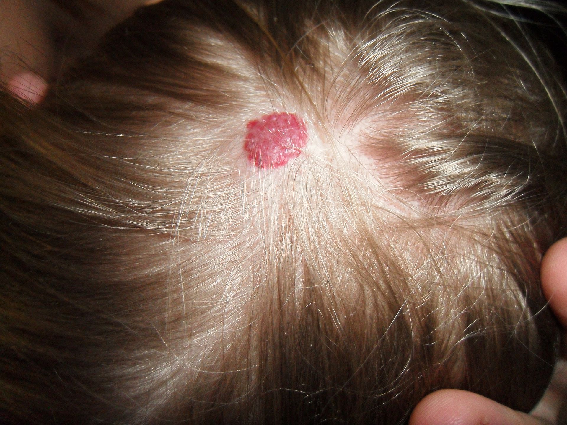
Capillary haemangioma (strawberry naevus)
This is a common benign tumour in infancy of the capillary system.
– Usually found on the face, scalp and back and seen in about 10% of Caucasian infants
– It can bleed, ulcerate and also rarely obstruct the visual fields or airway
Appearance – Presents as red, raised, multilobed mark on the skin
Septal Haemangiomas
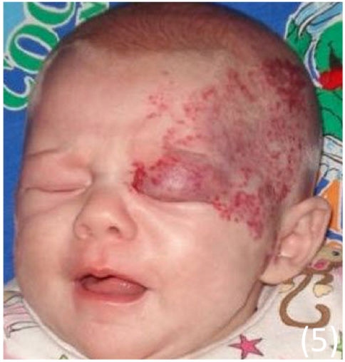
– more serious, occur at a younger age and often 10x larger Facial segmental haemangiomas are associated with PHACE syndrome
– Posterior fossa abnormalities
– Haemangiomas
– Arterial abnormalities
– Cardiac abnormalities
– Eye abnormalities
The haemangioma also causes vascular steal from other developing tissues, giving other abnormalities.
Management
– We only treat certain hemangiomas which present specific risks. These include:
– Ulceration
– Airway obstruction
– Periocular haemangiomas – these often compromise visual axis and development of binocular vision
1st line treatment = Oral propranolol (b-blocker) in 1st year
– Alters expression of growth factor
– Gives increased apoptosis
– Increased constriction of blood vessels
Vascular malformations
This refers to an error in the development of vascular embryological tissue.
– These are present at birth and grow slowly and proportionately with the child:
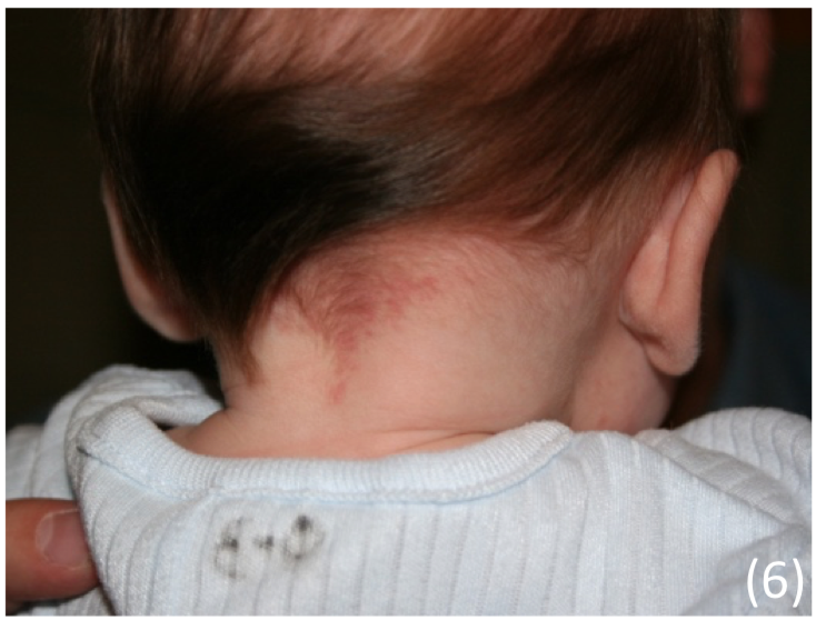
Stork mark (naevus simplex)
This occurs due to a (transient) capillary malformation giving marks which become more prominent with crying. Occurs in 40% of newborns
Appearance
– A flat, pink macule which occurs on forehead, eyelids and back of neck
Management
– Lesions usually fade spontaneously within 2 years
Port wine stain (PWS)
This is a type of permanent capillary malformation which is present at birth
Appearance
– Pink macule with a midline cutoff
– It occurs anywhere including the face
– Often darken and become elevated with time
Management
– No cure available
– Laser therapy can be used to reduce the colour
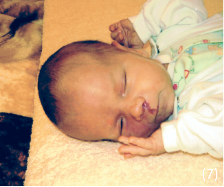
N.B. A PWS in the frontal placode is associated with Sturge Weber Syndrome:
– This is a rare congenital neurological and skin disorder, due to genetic mutation in gene GNAQ
– Causes overabundance of capillaries around ophthalmic branch of CN V + malformation of blood vessels
Symptoms:
– Port wine stain –> due to abnormal development of vascular tissue
– Seizures –> due to vessel abnormality to the leptomeninges
– Glaucoma –> due to vessel abnormality around the eyes
Management:
– Treated on a symptomatic basis e.g. ophthalmology for glaucoma, anti-epileptics for seizures
– Cutaneous mark – partially treated by laser therapy
Sources
Image 1: Gzzz / CC BY-SA (https://creativecommons.org/licenses/by-sa/4.0)
Image 2: Arya S, Jain M, Bunkar M, Takhar R / CC BY-SA (https://creativecommons.org/licenses/by/4.0)
Image 3: User:Zeimusu / Public domain
Image 4: Cbheumircanl / Public domain
Image 5: Haggstrom AN, Garzon MC, Baselga E, Chamlin SL, Frieden IJ, Holland K, et al. Risk for PHACE syndrome in infants with large facial hemangiomas. Pediatrics 2010;126:e418–26.
Image 6: Abigail Batchelder / CC BY -SA (https://creativecommons.org/licenses/by/2.0)
Image 7: ArturroD / CC BY-SA (https://creativecommons.org/licenses/by-sa/3.0)