Back to: Gastro-Intestinal
Biliary Conditions
Biliary Colic
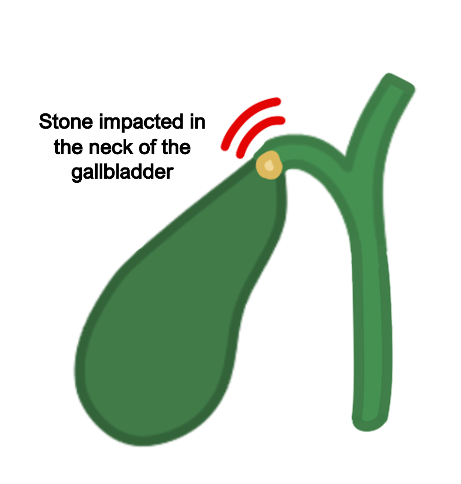
This is writhing right upper quadrant pain which occurs due to the gallbladder contracting to clear a stone stuck in the cystic duct or gallbladder neck.
Pain usually occurs after a fatty meal when the gallbladder contracts to release bile.
If left untreated this can lead to inflammation causing acute cholecystitis.
Symptoms
Right upper quadrant pain (can radiate to the right shoulder and scapula)
Nausea and vomiting
No fever or jaundice
Key tests
Ultrasound to detect stone
LFTs are usually within normal limits
Management
Small stones can pass spontaneously with resolution of the symptoms
If persistent, may require a cholecystectomy
Mirizzi syndrome
This is when a stone in the gallbladder neck causes compression of the CBD or common hepatic duct, resulting in an obstructive jaundice.
Usually stones in the gallbladder are asymptomatic, but this is a special case where it can compress the common bile duct, leading to obstruction of bile flow.
This increases the risk of cholecystitis and cholangitis due to CBD obstruction.
Surgery is difficult as Calot’s triangle can be obliterated, increasing the risk of common bile duct damage.
Acute Cholecystitis
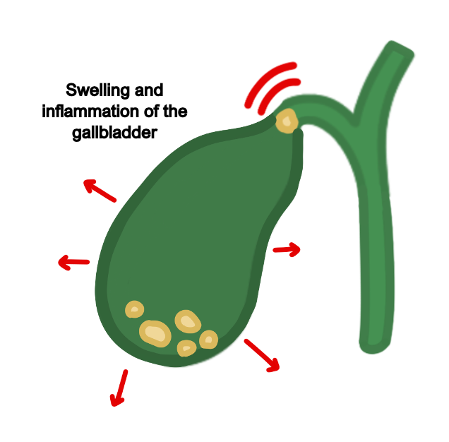
This refers to acute inflammation of the gallbladder.
It is usually secondary to a stone in the cystic duct which causes dilation, bacterial (E. coli) growth and inflammation.
Symptoms
RUQ pain, nausea/vomiting, fever
No jaundice
Murphy’s sign positive – palpating the RUQ causes pain during inspiration, as the inflamed gallbladder touchers fingers
Key tests
Ultrasound shows stones
Blood tests show raised inflammatory markers (WBC, CRP) and raised alkaline phosphatase (from cystic duct damage)
Management
Provide analgesia and IV fluids
Broad spectrum antibiotics, e.g., co-amoxiclav
Once the infection settles, elective cholecystectomy
Choledocholithiasis
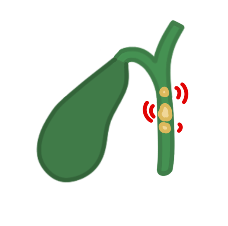
This is a stone in the common bile duct, which causes an obstructive jaundice
Symptoms
RUQ pain, nausea/vomiting and jaundice but no fever
Murphy’s sign negative as the gallbladder is not inflamed
If left untreated, this can lead to infection leading to ascending cholangitis
Key tests
Blood tests show raised ALP raised conjugated bilirubin
Urine dipstick shows elevated bilirubin
Imaging – ultrasound is gold standard
MRCP can be used if US is negative
Management
Removal of stone using ERCP followed by elective cholecystectomy
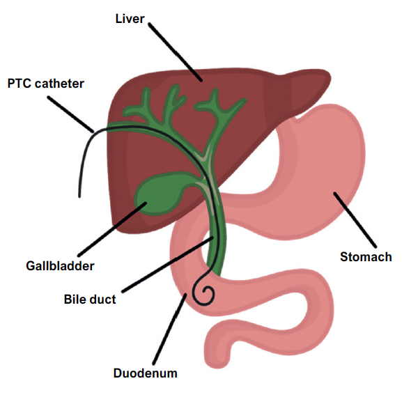
If ERCP is unsuccessful, percutaneous transhepatic cholangiography (PTC) can be done.
This is a tube which enters through the hepatic duct and allows the drainage of bile. It relieves pressure and can be used to remove the stone.
Ascending Cholangitis
This is a bacterial infection of the bile ducts, which often occurs after a stone gets stuck in the common bile duct.
It is usually due to an ascending infection secondary to enteric bacteria E. coli.
Symptoms
Charcot’s triad: RUQ pain, jaundice, fever
Reynold’s pentad: Charcot’s triad with hypotension and confusion
Key tests
Bloods show high ALP, raised bilirubin, raised inflammatory markers
Imaging – ultrasound and MRCP
Management
Sepsis 6 protocol with IV fluids with broad spectrum antibiotics
Removal of stone via ERCP, followed by elective cholecystectomy later
If ERCP unsuccessful, consider PTC or surgical exploration of the bile ducts
Gallstone Ileus
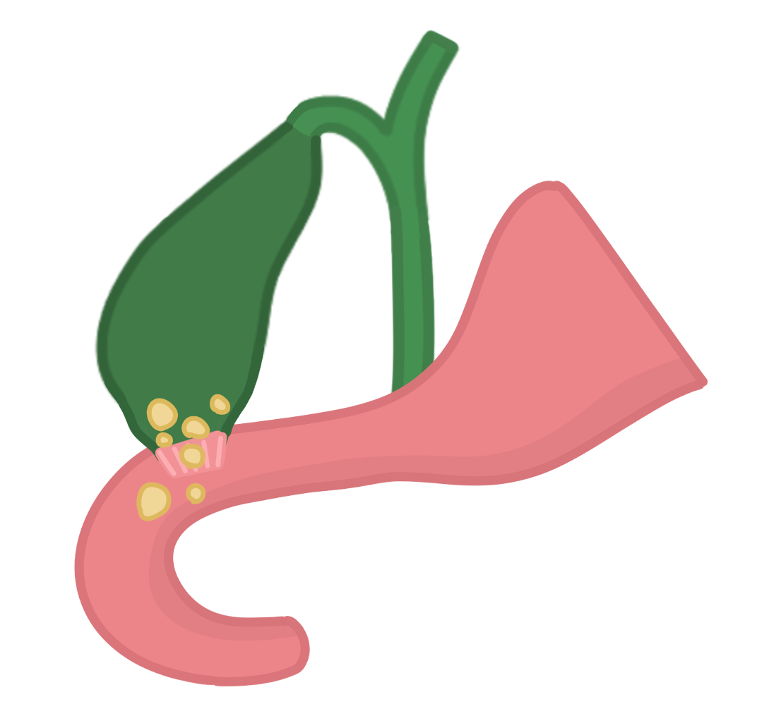
A condition caused by fistula formation between the gallbladder and duodenum.
This allows a gallstone to pass into the duodenum and cause small bowel obstruction, which commonly occurs near the distal ileum.
Duodenal obstruction is much rarer and known as Bouveret’s syndrome.
Symptoms
Usually preceded by history of cholecystitis that then gives obstruction
Bowel obstruction symptoms – nausea, vomiting, abdominal pain and distension
Key tests
Abdominal X-ray/CT scan shows small bowel obstruction with presence of pneumobilia and stone in right iliac fossa
Management
“Drip and suck,” IV fluids and decompression (NG tube) to treat bowel obstruction
Surgery may be required to remove the gallstone, with elective cholecystectomy
Cholangiocarcinoma
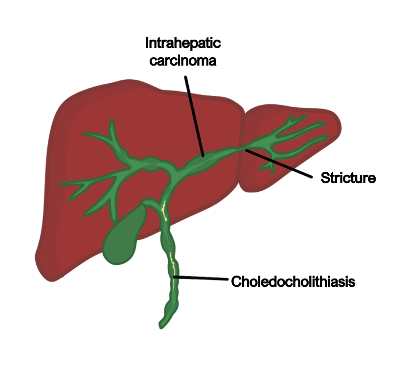
This is an adenocarcinoma arising from the epithelium lining the bile ducts.
The major risk factor is primary sclerosing cholangitis as well as hepatitis B/C.
Symptoms
Abdominal pain, itching and weight loss
Palpable RUQ mass (Courvoisier’s sign) due to gallbladder dilation
Obstructive jaundice, with changes in the colour of stool and/or urine
Key tests
Bloods show high ALP, bilirubin, raised cancer markers CA 19-9, CEA, CA-125
CT/MRI and MRCP show biliary tree dilation.
Biopsy may be required for histology
Management
Mainly involves palliative chemotherapy with biliary stenting

