Urea and Electrolytes (U&Es)
This blood test is the simplest biochemical measure of assessing renal function.
By looking at the plasma concentration of creatinine and urea, this allows you to estimate the glomerular filtration rate (GFR) to see if the kidneys are effectively filtering the blood.
Creatinine
This is a breakdown product of creatine phosphate in skeletal muscle.
It is freely filtered, not reabsorbed, and slowly secreted.
GFR is inversely proportional to the plasma creatinine concentration.
As creatinine is proportional to muscle mass, the result requires adjustment using the Modification of Diet in Renal Disease (MDRD) (or equivalent GFR estimation equations) equation to calculate the effective GFR (eGFR).
Creatinine is affected by pregnancy, muscle mass and diet (e.g., eating red meat)
Urea
Urea is a waste product which is formed from the breakdown of proteins.
Urea is usually excreted by the kidneys, so a decrease in kidney function will cause urea levels to rise.
The serum urea: creatinine ratio can be used to ascertain the origin of an acute kidney injury.
Although serum urea levels are used to assess renal function, changes in urea levels may also indicate other pathologies.

Sodium
The serum sodium concentration should be the 136-145mM. Both high and low levels of sodium are associated with different causes and symptoms.
Hyponatraemia
This is a low concentration of plasma Na+. As the concentration depends on both sodium levels and water volume, hyponatreamia can either be due to a lack of sodium ions or too much water.
Causes
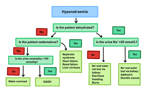
The main way to distinguish the causes is first assessing if the patient is dehydrated or full of water.
If dehydrated, this suggests the primary problem is losing Na+ ions, and water indirectly follows as a result.
Kidney etiology – Addison’s disease or renal failure
Outside kidneys – Burns, diarrhea, small bowel obstruction
If not dehydrated, this suggests that there is excessive relative water retention occurring, causing the plasma to become more diluted.
If patient has oedema (hypervolaemic), suggests kidney, liver, heart failure
If no oedema (euvolemic), either SIADH or water overload

Central Pontine Myelinosis
If the sodium level is corrected too quickly causes central pontine myelinosis
– This is rapid demyelination of the pons
– Gives lethargy, confusion, seizures and locked in syndrome (paralysis of limbs) with coma
– Therefore, aim to increase his sodium by <2 mM/hour, and definitely no more than 10 mM/day.
Hypernatremia
This usually occurs due to water loss in excess of Na+ loss (high osmolality)
Causes
DEHYDRATION
DIABETES insipidus
DOCTORS: incorrect IV fluid replacement (excessive saline)
DIURETICS
DIARRHOEA
DISEASE (e.g. renal)
Symptoms
Thirst
Weakness and lethargy
Irritability, confusion and coma
Management
If in coma – glucose 5% IV slowly guided by fluid balance and sodium levels
Again, be careful when correcting levels as quick correction can lead to cerebral oedema raising ICP
Potassium
The Potassium concentration in the blood should be between 3.5 and 5.3mM.
Hypokalaemia
This hyperpolarizes the cell and reduces the excitability of nerve and muscles, causing muscular weakness which leads to muscular paralysis and cardiac arrhythmias
Symptoms
Muscle weakness, with hypotonia and hyporeflexia (decreases excitability of nerve + muscle)
Light-headedness
Arrhythmias and palpitations
Constipation
Key tests
ECG shows small or inverted T waves, long PR interval + prominent U wave
If person is on diuretics, raised HCO3- is best indicator that hypokalemia is long standing
Management
If mild, oral K+ supplement
If <2.5mM, IV KCl replacement with cardiac monitoring. (Max rate infusion = 10mM/hour)
Hyperkalaemia
This often due to renal failure where GFR drops below 20%
Raised potassium depolarises cells bringing them closer to threshold, increasing excitability of nerve and muscle and raising chances of cardiac arrhythmias.
Symptoms
Tachycardic irregular pulse,
Palpitations, chest pain and light-headedness
Key tests
ECG shows tall tented T waves, wide QRS (can lead to VF)
N.B. If patient is well but has a high K+ this can be due to artefact. This is caused by K+ leaking out of RBCs –> due to haemolysis, contamination, delayed analysis
Management
If single random high K+ reading, consider repeating the sample to ensure not an artefact.
Perform an ECG and treat if K+ >6.5mM or if there are visible ECG changes:
Calcium chloride/ or calcium gluconate IV, repeated if necessary
IV insulin in dextrose solution
Nebulized salbutamol (but avoid in tachycardias)
Disclaimer


