Persistent Cough in a Smoker
Try out this respiratory case and test your clinical knowledge. The answers are at the bottom.
Questions
A 59-year-old with COPD presents to their GP with a worsening cough. He says the cough has been going on for the last four weeks, is not productive but does keep him awake at night. He also reports that he has been passing urine more often but puts it down to anxiety.
Examination:
Faint wheeze bilaterally, good air entry
Observations:
SpO2: 95%
Temperature: 37.1
BP: 121/82
HR: 87
Q1: What is the differential diagnosis for his initial presentation?
Q2: How should the GP manage this patient (& why)?
The GP manages the patient appropriately. Some initial bloods and imaging are also performed:
Summary of Blood Results:
| Test | Result | Reference Range |
| FBC | 120 | 135 – 180 g/l |
| WCC | 4.3 | 4 – 11 x 109/l |
| Plts | 161 | 150 – 400 x 109/l |
| MCV | 87 | 82 – 100 fl |
| Na+ | 134 | 135-145 mmol/l |
| K+ | 4.5 | 3.5 – 5 mmol/l |
| ALP | 230 | 30 – 100 umol/l |
| PTH | 1.1 | 1.6 – 6.9 pmol/l |
| Ca2+ | 2.9 | 2.1 – 2.6 mmol/l |
| Phosphate | 0.6 | 0.8 – 1.4 mmol/l |
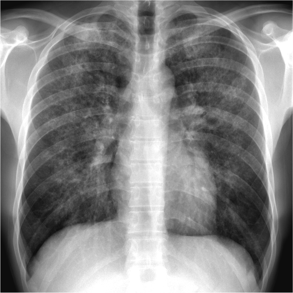
Chest X-Ray:

Q3: Comment on the blood results and CXR. What is the likely diagnosis?
Q4: What other complications can develop from this condition?
Answers
Reveal the answers
- Exacerbation of COPD – unlikely due to lack of productive cough and lack of fever
- Pneumonia – unlikely due to lack of productive cough and lack of fever
- PE – unlikely due to time-course and lack of risk factors
- Sinusitis – lack of post-nasal drip, stuffy nose, facial pain
- Asthma exacerbation – no history of atopy or asthma
- Bronchiectasis – possible diagnosis
- Asbestosis – no history of occupational exposure
- Idiopathic pulmonary fibrosis – possible diagnosis
He is over 40 and is presenting with a new-onset cough on a background of significant smoking history (COPD diagnosis) – he meets the 2-week wait referral criteria for a CXR
Diagnosis: Squamous cell carcinoma of the lung
Bloods:
- Normocytic anaemia – likely increased demand for oxygen
- Mild hyponatremia – not significant
- High ALP – ectopic PTHrp released from cancer stimulates osteoclasts, leading to increased bone resorption and hence a rise in ALP
- Low PTH – endogenous PTH is suppressed due to the elevated serum calcium
- High calcium – Hypercalcaemia due to the effects of PTHrp on bone and kidneys
- Low phosphate – PTHrp stimulates phosphate excretion from the kidney
Chest X-Ray:
- Tracheal deviation to R side
- ‘Sail’ sign in R lower zone – indicates R lower lobe collapse
- Large collapse like this indicates major airway e.g. large segmental bronchus occlusion
- The usual central location of squamous cell carcinoma supports this finding
- Hyperthyroidism (Ectopic TSH)
- Hypertrophic pulmonary osteoarthropathy
- Metastatic disease
To get more information about the conditions mentioned in this case including diagnosis and management, have a look at our free respiratory notes on In2Med. Written by medical students, we have pitched them just at the right level to help you ace your exams.
Sources
Image 1: Radiopedia https://bityl.co/6B9g
University of Cambridge
About The Author
Disclaimer


