Hip X-Rays
Check out these examples of hip conditions. Have a read of the first example where we go through how to present this plain film.
Practice with the remaining examples and click on the box to reveal the diagnosis.
Example 1
Take a look at the following example. Let us go through how we would systematically analyse this and the diagnosis.
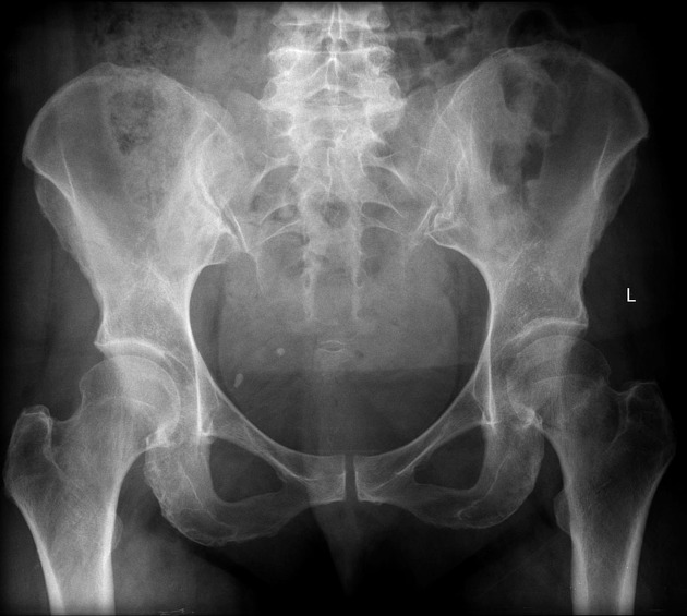
Analysis and Diagnosis
D – This is a … X-Ray taken on ….., of the following patient….. Is there a previous X-ray to compare to
R – Commenting first on the quality, it is not rotated, there is adequate field of view, the projection is AP and lateral and it is adequately exposed as I can see the bones clearly
“On initial inspection, there appears to be a fracture of the neck of femur, but I will proceed to go through it systematically.”
B – Looking at the bones first, there is a subcapital fracture in the neck of femur. It is a closed, simple, transverse fracture. The fracture line extends through the junction of the head and neck of femur. There is no translation, rotation or shortening. There are no other fractures.
There are no obvious dislocations and the no abnormality in the texture of the bones
D – There are no degenerative changes in the joint spaces
E – There are no other man-made objects or signs of orthopaedic surgery.
In summary, this film shows an intracapsular fracture of the proximal femur.
Diagnosis
Neck of Femur Fracture
Example 2
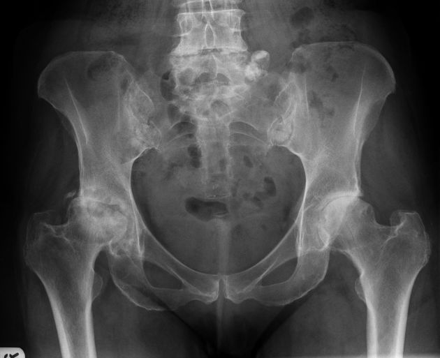
Diagnosis
Osteoarthritis of the Hip (right)
Example 3
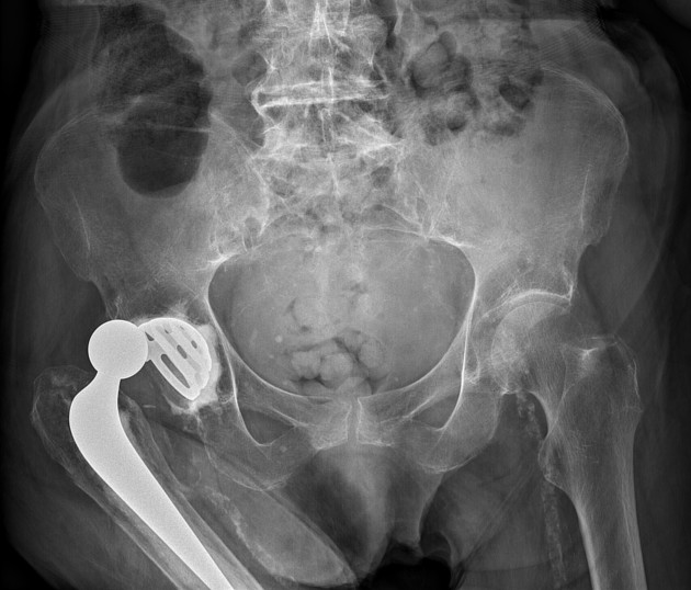
Diagnosis
Total Hip Replacement
Example 4
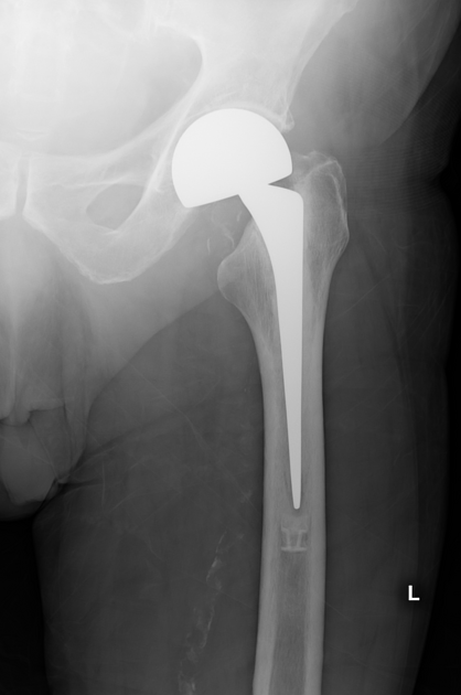
Diagnosis
Hemiarthroplasty
Example 5
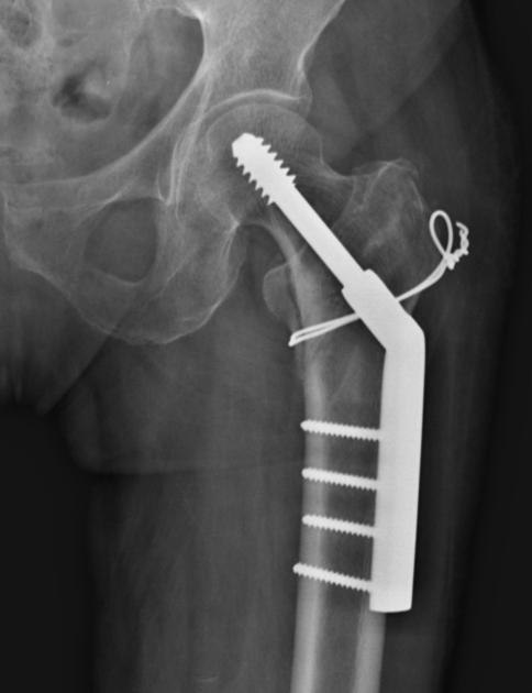
Diagnosis
Dynamic Hip Screw
Check out the following pages to see more examples of fractures and dislocations and how you would systematically analyse the X-Rays.
Sources
Image 1: Gaillard, F. Impacted subcapital fracture. Case study, Radiopaedia.org. https://doi.org/10.53347/rID-2717
Image 2:Feger, J., Niknejad, M. Osteoarthritis of the hip. Reference article, Radiopaedia.org. https://doi.org/10.53347/rID-81200
Image 3: Hacking, C., Qureshi, P. Total hip arthroplasty. Reference article, Radiopaedia.org. https://doi.org/10.53347/rID-37780
Image 4: Hacking, C., El-Feky, M. Hip hemiarthroplasty. Reference article, Radiopaedia.org. https://doi.org/10.53347/rID-37709
Image 5:Hacking, C., Bell, D. Dynamic hip screw. Reference article, Radiopaedia.org. https://doi.org/10.53347/rID-37374
Disclaimer


