Tachycardia
This is defined as a heart rate of > 100 bpm. Whilst it can be normal physiological response to increased demand (e.g., exercise), there are many arrythmias which also give rise to tachycardias.
Atrial fibrillation
This is a chaotic irregular atrial rhythm with an atrial rate which can exceed 400 bpm.
Small sections of atria are activated individually, resulting in the atrial muscle quivering instead of contracting in a synchronised manner.
The AV node allows intermittent impulses, which leads to an irregular ventricular contraction rate.
It is usually asymptomatic as the cardiac output only drops by about 10%, but it can lead to palpitations and haemodynamic compromise if severe.
Types
Atrial fibrillation can be classified into sub-categories:
First detected episode – this is the first time the patient experiences this arrythmia
Recurrent – this is when patient has multiple episodes of AF (in 2 subtypes):
Paroxysmal AF – when episodes of AF terminate spontaneously, usually < 7 days
Persistent AF – when episodes are not self-terminating and usually last > 7 days
Permanent AF – where the AF is unresponsive to cardioversion or other treatment
Causes
Many cases due to atrial hypertrophy/dilatation
Risk factors include hypertension, age, obesity, heart failure or mitral valve disease
Ischaemic heart disease, idiopathic and infection
Decreased K+, decreased Mg2+, caffeine, hyperthyroidism and regular alcohol use
Symptoms
Can be asymptomatic
Chest pain, palpitations, hypotension
Irregularly irregular pulse – this is caused by the fact that the AV node only allows intermittent pulses
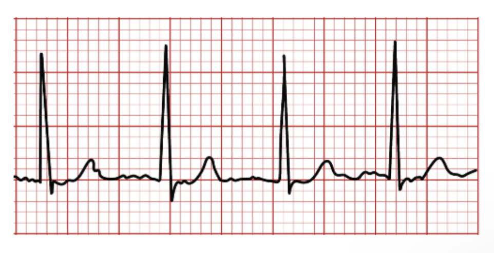
ECG Features
P waves absent – replaced by f waves.
Rhythm is irregularly irregular
Patient usually tachycardic
Normal narrow QRS complexes
Management
If hemodynamically unstable, electrical DC cardioversion with optional amiodarone
If hemodynamically stable, management consists of a rate or rhythm control strategy depending on the patient
Rate control
Here, it is accepted that the pulse will be irregular, but we give medications in order to slow the heart rate down.
1st choice is a beta-blocker (e.g., atenolol) or a rate-limiting Ca2+ blocker (diltiazem)
If not effective, dual therapy with drugs or addition of digoxin
Rhythm control
This attempts to get the patient back into normal sinus rhythm
Can use electrical cardioversion or drugs (Amiodarone or flecainide)
If onset is > 48 hours, it is advised to give anticoagulation for at least 3 weeks before performing cardioversion, unless the patient is haemodynamically unstable.
| Rate Control Preferred | Rhythm Control Preferred |
| Patient is over age 65 years | Patient is younger than 65 years old |
| Asymptomatic | Symptomatic |
| AF is permanent | First detected episode of AF |
| No reversible cause | Due to a reversible cause e.g pneumonia |
| Due to myocordial infarction | Patient has symptoms of heart failure |
In addition, the main risk of AF is embolic stroke.
The CHA2DS2-VASc score is used to assess embolic stroke risk. Warfarin/NOACs is given to patients with a score of 2 or more (+ some men with score of 1):
CHA2DS2-VASc Score
| Variable | Points Awarded | |
| C | Congestive heart failure | 1 |
| H | Hypertension (BP > 140/90 mmHg) | 1 |
| A2 | Age over 75 years | 2 |
| Age over years | 1 | |
| D | DIabetes | 1 |
| S2 | Strike or TIA in medical history | 2 |
| V | CardioVascular disease | 1 |
| S | sex = female (higher risk due to raised oestrogen) | 1 |
Atrial flutter
This is a supraventricular tachycardia characterized by a fast atrial rate of around 200-400bpm.
The AV node again behaves intermittently, usually impeding many impulses
Crucially however, this delays impulses consistently and so the ventricular rate will probably be regular
Causes
Found in conditions that enlarge atrial tissue e.g., mitral disease, acute MI
Symptoms
Can be asymptomatic or cause chest pain, palpitations, hypotension
Clinical significance is determined by conduction ratio – number of impulses through node.
If it drops too low, cardiac output will be compromised –> can cause syncope, angina etc.
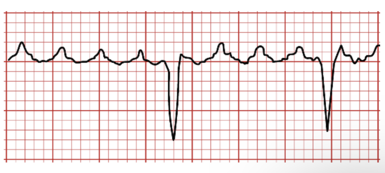
ECG Features
P waves lose distinction due to rapid rate and blend together
They give a “saw-toothed” appearance and are called flutter (F) waves.
Normal QRS waves
Management
Same management as atrial fibrillation. It is generally more difficult to rate-control than AF and therefore cardioversion is used more frequently.
If recurrent, radiofrequency ablation of the abnormal re-entrant circuit via ablating the cavo-tricuspid isthmus (CTI) is > 90% successful.
Supraventricular Tachycardia (SVT)
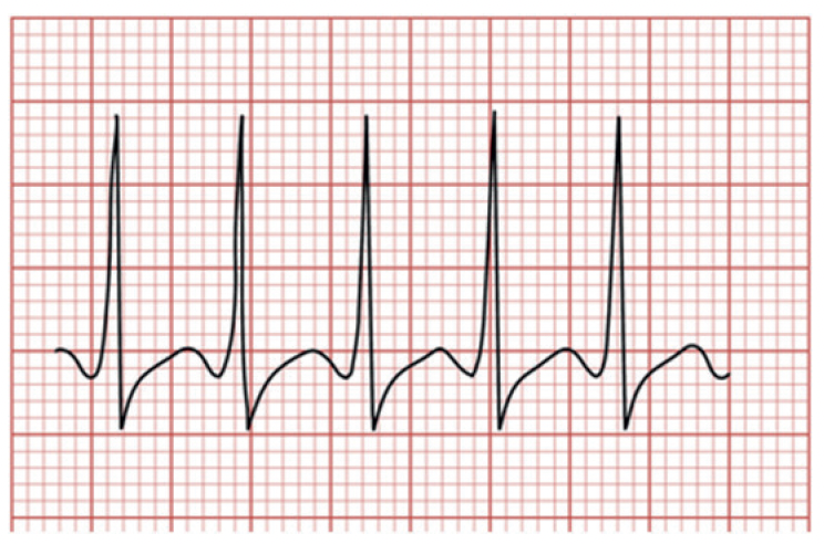
An episodic tachyarrhythmia which has an unpredictable onset and termination.
It is typically an atrioventricular nodal re-entry tachycardia (AVNRT).
It leads to acute attacks of palpitations with symptoms which may then spontaneously resolve.
Symptoms
Episodes start and end suddenly but can be asymptomatic
Palpitations with chest pain
Sudden onset dyspnoea and feeling faint
Management
1st line is vagal maneuverers to increase parasympathetic tone slowing the heart rate, e.g., Valsalva manoeuvre, carotid sinus massage
If unresolving, IV adenosine 6 mg. If still unsuccessful, this can be repeated at a dose of 12 mg and third dose of 18 mg
If persistent, electrical cardioversion can be used
In the long term, these patients will likely need radio-frequency ablation to ablate the abnormal electrical circuit
Wolff-Parkinson White (WPW) syndrome
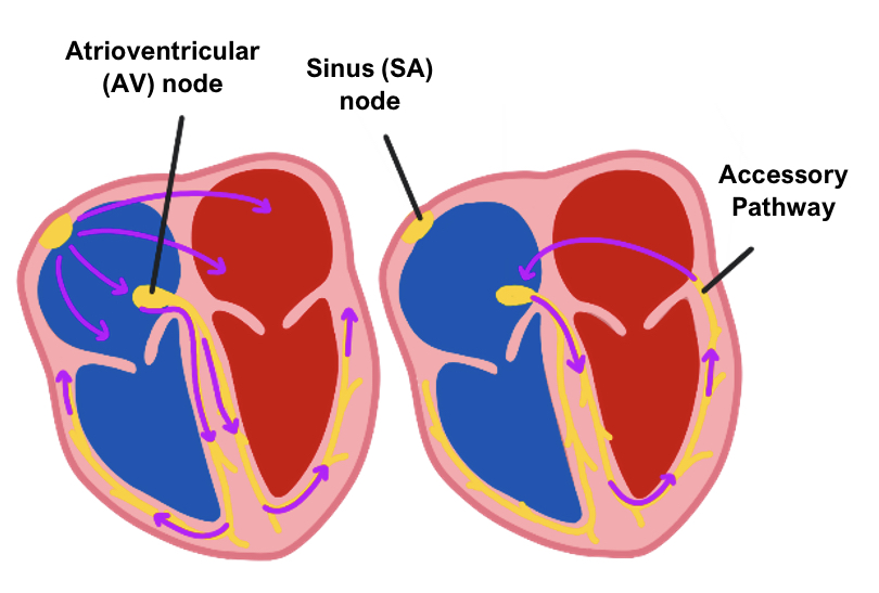
This is a condition in which there is an abnormal accessory conduction pathway between the atria and ventricles.
This means that the electrical impulse can bypass the delay at the AV node causing a fast heart rate.
As the accessory pathway does not slow conduction, it means atrial fibrillation can rapidly lead to ventricular fibrillation causing sudden death.
Any medication that blocks the AV node can potentiate the arrhythmia by promoting antegrade conduction down the accessory pathway.
Associations
HOCM
Mitral valve prolapse
Ebstein’s anomaly
ECG Features
Short PR interval
Wide QRS complex with a slurred upstroke = “delta wave”
Type A (left-sided pathway) gives right-axis deviation
Type B (right sided pathway) gives left- axis deviation
Management
If hemodynamically unstable, electrical cardioversion
Medical management involves medications like amiodarone and procainamide
Definitive management is radiofrequency ablation of the accessory pathway
Ventricular Tachycardia (VT)
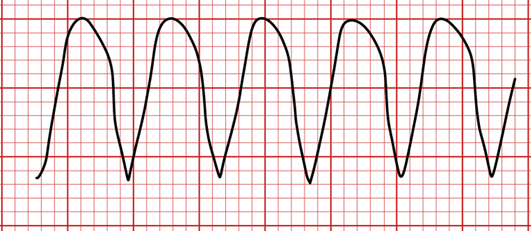
This is a condition which occurs when the ventricular rate exceeds 100 bpm.
It is an unstable rhythm and can quickly progress to ventricular fibrillation.
As the rate originates in the ventricles, it causes broadening of the QRS complex.
On ECG, P waves are not seen as the ventricles overshadow the atrial impulse.
Monomorphic VT
This is a broad complex tachycardia with a ventricular rate > 100 bpm.
It is most commonly due to ischaemia of the heart (e.g., MI) or a scar from previous ischaemic events providing a substrate for the VT.
Causes
Most commonly due to ischaemic to the heart (e.g. MI)
ECG features
Atrial rhythm/rate can’t be determined.
Rhythm regular and rapid >100bpm.
QRS is wide(>0.12s) and increased amplitude
Management
If patient is pulseless, treat as cardiac arrest
If hemodynamically unstable (BP < 90 mmHg, chest pain), electrical cardioversion
If hemodynamically stable, 1st line is IV amiodarone, 2nd line is lidocaine
If not due to an acute MI, the patient will likely need an implantable cardioverter-defibrillator (ICD)
Polymorphic VT (Torsades de pointes)
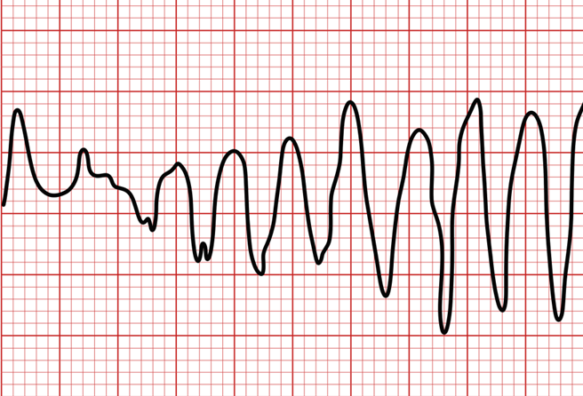
This is a type of VT which is associated with having a long QT interval.
The rate is again generated by the ventricles, but it does not give a consistent welldefined broad complex tachycardia like monomorphic VT.
Causes
Due to factors which increase the QT interval
Drugs (amiodarone, TCAs, erythromycin)
Hypothermia
Hypokalemia/Hypocalcaemia
ECG features
Hallmark is QRS complexes that rotate about the baseline, deflecting down and up for several beats.
Tachycardia (rate >100bpm)
Management
Intravenous infusion of magnesium sulfate is usually effective. Patients may require cardioversion or pacing.
Disclaimer
