Bradycardias
Bradycardia is defined as a heart rate of fewer than 60 beats per minute.
Bradycardia can be normal in some people, but can also lead to haemodynamic compromise, leading to the development of symptoms.
Symptoms
Hypotension (< 90mmHg)
Syncope
Heart failure
Management
First-line is atropine 500 mcg IV – this can be repeated up to a maximum of 3 mg
If no improvement, use transcutaneous pacing
If persistent, give isoprenaline/adrenaline infusion titrated to response
Long term management involves insertion of a pacemaker
Atrioventricular heart block
This is an abnormality in the normal electrical conduction of the heart, which leads to delayed transmission of the impulse, resulting in a slow heartbeat.
Atrioventricular heart block results from the interruption of impulse conduction from the atria to the ventricles.
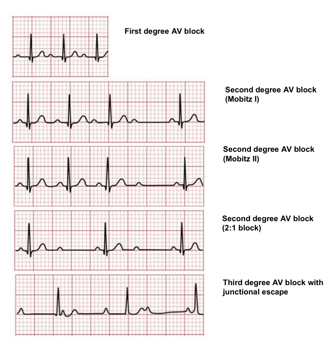
AV blocks are classified according to their electrographic appearance, which gives a clue as to the location.
1st degree heart block
This is when impulses from the atria are consistently delayed by a certain amount at the AV node.
It can be a normal variant and is often asymptomatic.
Causes
Idiopathic
Digoxin
Coronary artery disease (CAD)
Lyme disease
Symptoms
Often asymptomatic as cardiac output not affected
ECG Features
The PR interval is prolonged (> 0.20 s), but it is the same in each beat.
There are no missed beats.
2nd degree heart block: Mobitz type I
This is when each successive impulse is delayed more and more by the AV node.
This continues until an impulse fails to be conducted from ventricles, resulting in a missed beat, which then restarts the cycle.
This can also be a normal variant, especially in very fit people at rest.
It is not generally dangerous, but pacing can be considered if it causes symptoms such as dizziness or it is affecting the quality of life.
Causes
Idiopathic
CAD
Inferior MI
Usually temporary due to medication (B-blockers + digoxin)
Symptoms
Often asymptomatic but might show light-headedness or hypotension
ECG Features
PR interval is more and more prolonged each beat, until you miss a beat completely
This is called the Wenckebach phenomenon
2nd degree heart block: Mobitz type II
This is when impulses from the SAN are intermittently not conducted to the ventricles, usually (75%) distal to the bundle of His.
Due to the distal ocation of the conduction defect, the QRS often appears broad.
Mobitz type II requires permanent pacemaker implantation as there is a high risk of progression to complete heart block and sudden cardiac death.
Causes
Severe CAD
Anterior wall MI
Changes in the conduction system (structural damage)
Symptoms
Dyspnea
Fatigue
Light-headedness
Pain
Hypotension
ECG Features
Constant PR interval but many P waves are not followed by QRS
Ratio ranges from 2:1 to 4:1
3rd degree heart block
This is when impulses from SAN are completely blocked from reaching the ventricles.
The site of the block largely determines the ECG characteristics.
An escape rhythm from near to the AV node causes a relatively narrow QRS and higher rate (sometimes above 40 bpm).
A more distal block results in a broader ventricular escape rhythm, a lower heart rate and pacing is even more urgent.
Causes
Inferior wall MI
Congenital
Aortic valve calcification
Digoxin toxicity
Symptoms
Dyspnea
Fatigue
Light-headedness
Pain and hypotension
ECG Features
No relation between P waves and QRS waves, 2 independent rhythms present
Bundle Branch Block (BBB)
In the heart, the two bundles of His conduct electrical activity to the ventricles.
In BBB, either the left or right bundle fails to conduct impulses to the ventricles.
This means that the wave of depolarisation must pass via cell-to-cell conduction, which is much slower than along the specialised bundle branch cells.
This prolongs the contraction rate which results in widening of the QRS complex.
This can slow the heart rate down causing bradycardia, impairing the cardiac output.
It leads to the development of similar symptoms to AV heart block (hypotension, lightheadedness, chest pain etc.).
BBB can occur further down the left bundle, which is divided into an anterior and posterior fasciculus.
A branch block that occurs here is known as a hemiblock.
Right Bundle Branch Block
This is when the right bundle is blocked.
It is important to know that this can be a normal variant (seen more in the elderly)
Causes
Idiopathic (normal variant if isolated and asymptomatic)
Conditions which increase pressure in right side of heart – Pulmonary embolism, cor pulmonale
Right ventricular hypertrophy – after a Myocardial infarction, cardiomyopathy
Atrial septal defect
ECG Features
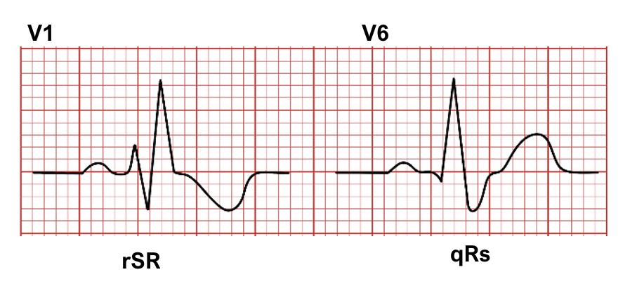
QRS >0.12s
MaRRoW sign on ECG
“RSR” pattern in Lead V1 (M)
“qRS” pattern in V6 (W)
Also may see inverted T waves in V1-3 or V4 and a wide slurred S wave in V6
Left Bundle Branch Block
This is a condition which describes when the left bundle is blocked giving QRS > 0.12s
This never occurs normally and is always considered pathological
Causes
Usually caused by ischaemic heart disease
New onset LBBB may be a sign of myocardial infarction in patients
Conditions which increase pressure in left side of heart – hypertension, aortic stenosis
ECG Features
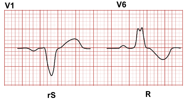
QRS >0.12s
WiLLiaM sign on ECG
“QRS” pattern in Lead V1 (W)
“RSR” pattern in V6 (M) – WiLLiaM
Bifascicular Block
This is a combination of RBBB and left anterior or posterior hemiblock
Trifascicular Block
This is a combination of bifascicular block accompanied with 1st degree heart block
Disclaimer

