How an ECG works
The ECG uses electrodes placed on the skin to measure the electrical activity of the heart.
The standard ECG consists of 10 electrodes, which provide a 12-lead output.
There are 6 electrodes placed on the chest and 1 on each limb.
One of these is a ground electrode to prevent electrical interference
The leads work by measuring electrical activity between two electrodes:
Depolarisation travelling in the direction of the positive electrode = positive (upwards) deflection on ECG
Depolarisation travelling away from positive electrode = negative (downwards) deflection on ECG
Limb Leads
These leads are bipolar leads which are placed on the arms and legs:
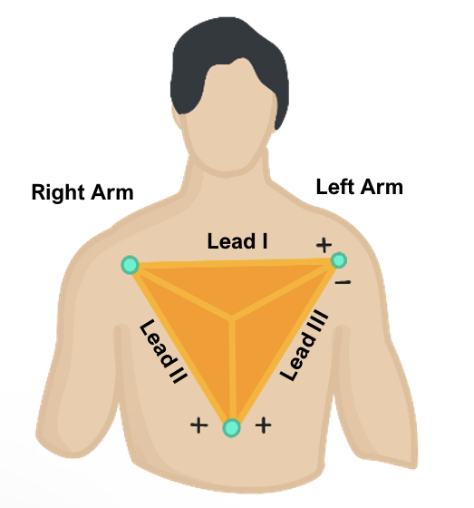
Lead I captures current moving form right to left: -ve electrode on right arm and +ve on left
Lead II has –ve electrode of right arm and +ve on left leg: therefore, produces the positive, high voltage deflections resulting in tall P, R and T waves.
Lead III has –ve electrode on left arm and + on left leg: useful for detecting changes in inferior wall
Using these three leads, they form Einthoven’s triangle around the heart and provide a frontal plane view.

Augmented leads
These are called augmented as the small waveforms that would appear are increased in amplitude. They are all positive electrodes and the negative electrode is the centre of the heart.
They also provide a view of the heart’s frontal plane.
Lead aVR – the positive electrode is on the right arm. This captures current moving from centre to right
Lead aVL – positive electrode on left arm. This captures current from the centre to left
Lead aVF – positive electrode on left leg. This captures current moving downwards
Using these 6 leads, you get a whole frontal view and so can work out the electrical axis of the heart.
Precordial leads
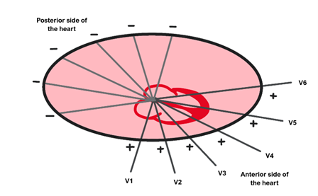
These leads are unipolar and placed across the chest to produce a view of the heart’s horizontal plane.
Lead V1 – right side of sternum at 4th intercostal space.
Lead V2 – left side of sternum at 4th intercostal space.
Lead V3 – between leads V2 and V4
Lead V4 – 5th intercostal space in mid-clavicular line
Lead V5 – 5th intercostal space in anterior-axillary line
Lead V6 – 5th intercostal space in mid-axillary line

Disclaimer


