Consolidation
Consolidation is one of the most important findings in a chest X-ray. Lung consolidation occurs when the air that usually fills the small airways in your lungs is replaced with something else. Consolidation occurs through accumulation of inflammatory cellular exudate in the alveoli and adjoining ducts.
The liquid can be pulmonary oedema, inflammatory exudate, pus, inhaled water, or blood (from bronchial tree or haemorrhage from a pulmonary artery).
Example 1
Take a look at the following example. Let us go through how we would systematically analyse this and the diagnosis.
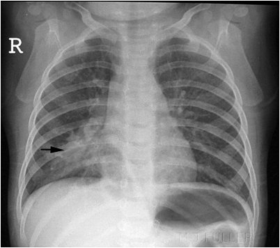
Analysis and Diagnosis
D – This is a Chest X-Ray taken on ….., of the following patient….. Is there a previous CXR to compare to
R – Commenting first on the quality, it is not rotated, there is adequate inspiration, the projection is posterior-anterior and it is adequately exposed as I can see the vertebral bodies clearly
“On initial inspection, there appears to be increased consolidation in the right middle lobe, but I will proceed to go through it systematically.”
A – Starting with the airways, the trachea is not deviated, and the carina is visible.
B – The pleural markings go all the way to the costal margin so there is no evidence of a pneumothorax. Going through the lung zones, there is increased opacification in the right middle lobe compared to the left, with obscuring of the right border of the heart. There is an air bronchogram present which is suggestive of middle love consolidation.
C – The heart is not enlarged. There is loss of the silhouette sign in the right border of the heart.
D – The hemidiaphragms are clearly visible and there is no blunting of the costophrenic angles. There is no free air under the diaphragm.
E – There are no foreign objects or bone fractures.
There is no abnormality in the review areas, including the apices, behind the lung.
Diagnosis
Right Middle Lobe Pneumonia
Example 2
Take a look at the following example showing consolidation. Click on the box to reveal the diagnosis.

Diagnosis
Right Upper Lobe Pneumonia
Example 3
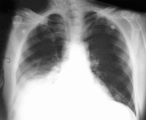
Diagnosis
Right lower lobe pneumonia
Example 4
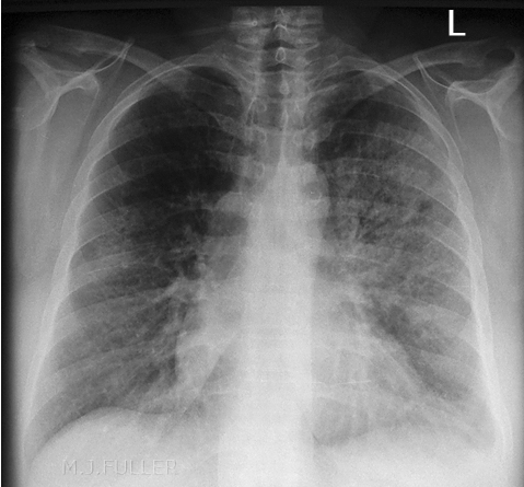
Diagnosis
Left Lung Lobar Pneumonia
Example 5
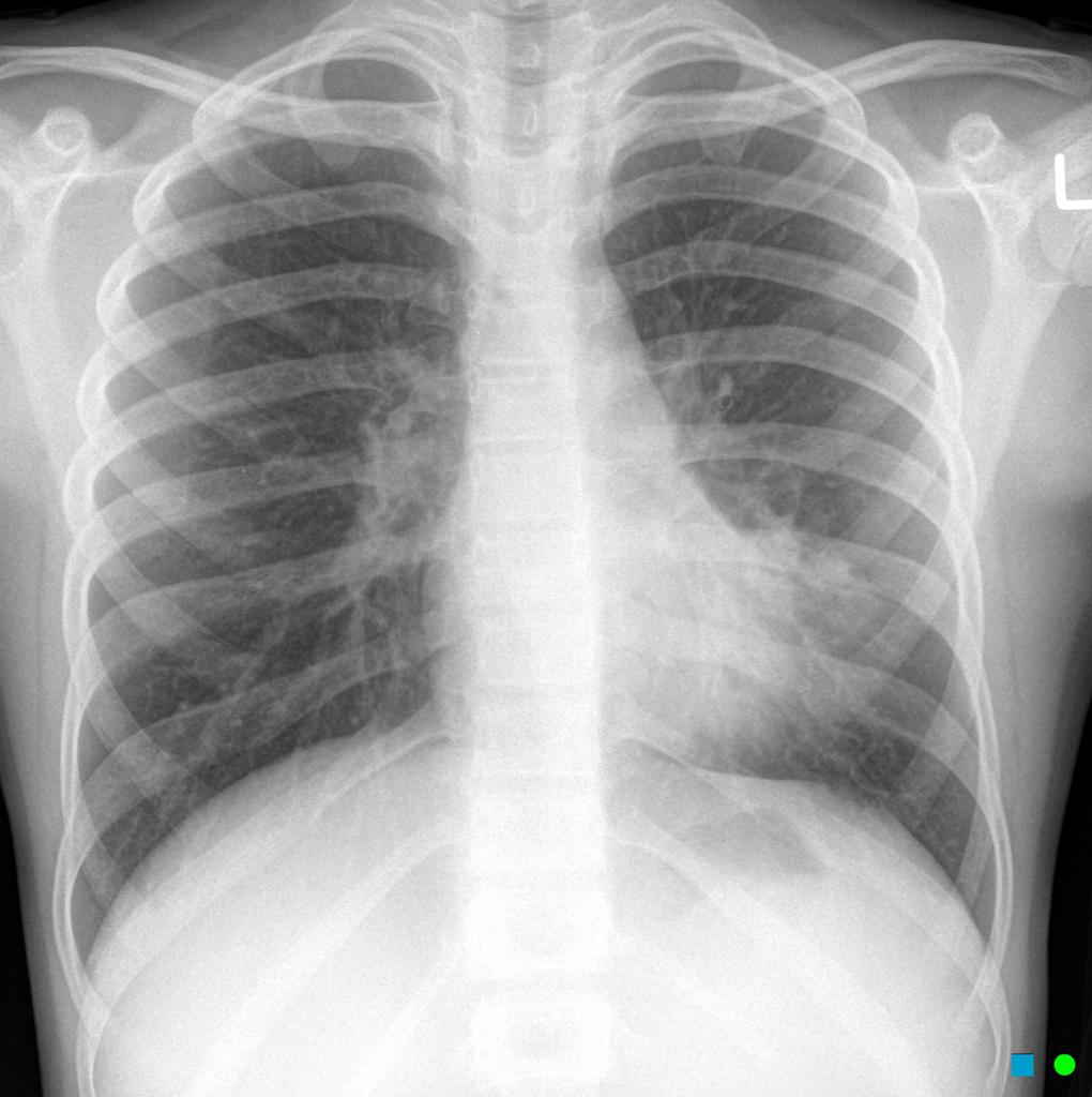
Diagnosis
Left Lingual Consolidation
Check out the following pages to see more examples of common pathologies seen on Chest X-Ray.
Sources
Image 1: http://www.wikiradiography.net/page/File:O2ZWd9HKVfas1Jyj3Ft1lw94915.jpeg#metadata
Image 2: http://www.wikiradiography.net/page/File:HtMjrgykbN2nPZ5L2sAU6g79538.jpeg
Image 3:http://www.wikiradiography.net/page/File:EA0lEXLTtJdDnFuMNQSYuA105014.jpeg
Image 4:http://www.wikiradiography.net/page/File:WqMK8cizeUcJWc4iIEcfaQ127018.jpeg
Image 5: Gaillard, F. Left lingula consolidation. Case study, Radiopaedia.org. (accessed on 11 Oct 2022) https://doi.org/10.53347/rID-7393
Disclaimer

