Masses and Raised ICP
Check out these examples of CT scans. See if you can identify the key finding and diagnosis.
We have analysed the first one for you. Click on the green box to reveal the diagnosis.
Example 1
Take a look at the following example.
Let us go through how we would systematically analyse this and the diagnosis.
Analysis and Diagnosis
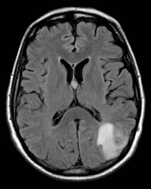
D – This is a CT Head taken on ….., of the following patient….. Is there a previous CT scan to compare to?
“On initial inspection, there is a suspicious lesion in the left parieto-occipital lobe, but I will proceed to go through it systematically.”
B – Looking at the film, there are no signs of an extra/subdural bleed and no intracranial bleeding.
C – The cisterns are clear and no sing of a subarachnoid haemorrhage.
B – The are no signs of a midline shift. There is a hyperdense area in the left parieto-occipital lobe. It has well defined borders and a ring of oedema around it.
V – The ventricles are not enlarged and there is no signs of an intraventricular bleed.
B – There are no obvious skull fractures.
In summary the key finding is a mass in the left parieto-occipital region.
Diagnosis
Tumour (metastasis)
Example 2
Take a look at the following example showing consolidation. Click on the box to reveal the diagnosis.
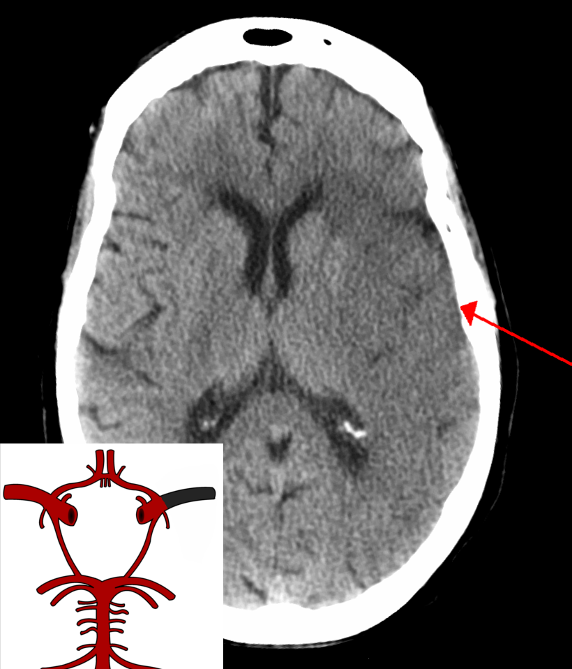
Diagnosis
Ischaemic Stroke
Example 3
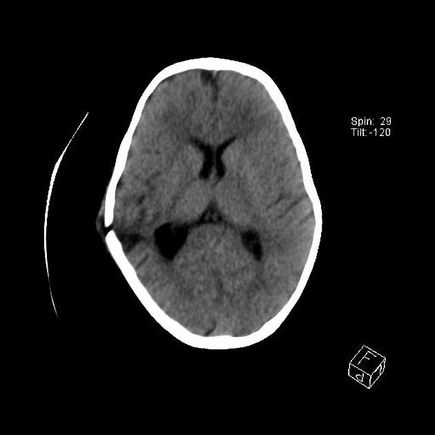
Diagnosis
Skull fracture
Example 4
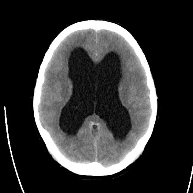
Diagnosis
Hydrocephalus
Check out the following pages to see more examples of common pathologies seen on CT head scans.
Sources
Image 1: http://www.wikiradiography.net/page/File:O2ZWd9HKVfas1Jyj3Ft1lw94915.jpeg#metadata
Image 2: http://www.wikiradiography.net/page/File:HtMjrgykbN2nPZ5L2sAU6g79538.jpeg
Image 3:http://www.wikiradiography.net/page/File:EA0lEXLTtJdDnFuMNQSYuA105014.jpeg
Image 4:http://www.wikiradiography.net/page/File:WqMK8cizeUcJWc4iIEcfaQ127018.jpeg
Image 5: Gaillard, F. Left lingula consolidation. Case study, Radiopaedia.org. (accessed on 11 Oct 2022) https://doi.org/10.53347/rID-7393
Disclaimer
