CT Head
Interpreting a CT/MRI Head Scan is an essential skill for any junior doctor. It is one of the most common first-line investigations that you will be asked to order, and can be used to investigate a variety of conditions.
To help you learn this skill, you can use the following Mnemonic “Blood Can Be Very Bad” – BCBVB.
By using this, you will be able to go through CT/MRI Head Scans in a systematic way to ensure you don’t miss any important details.
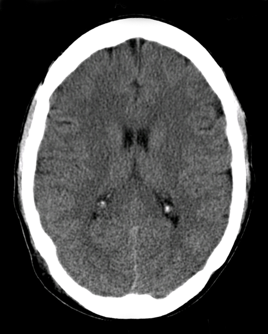
So if you are presented with a CT/MRI Head Scan like this, let’s see how to go through it.
Details
When starting with any CT/MRI Scan, the first thing to do is to confirm that it is of the right patient. This will involve checking for the following features.
- Confirm Name
- Confirm Age
- Confirm DoB
- Confirm Date and Time of Film
- General comment on adequacy of scan
- Is there anything I can compare the film to?
Many patient’s who have prolonged admissions or a background of neurological disease may have chronic CT head changes.
Therefore it is important to always ask whether there is a previous CT or MRI Scan that you can compare to.
This will help to identify whether any abnormality is longstanding or chronic.
State what you believe to be obvious pathology if you can then work through this systematically.
B – Blood
Now that you have confirmed patient details and commented on the film quality, you can start going through the CT Scan systematically.
The first part of the acronym (B) focusses on blood.
On a non-contrast CT, blood appears white, so it is fairly easy to spot large bleeds.
Ruling out a bleed is important if you suspect a thrombotic stroke, as bleeding is a contraindication to giving aspirin or other anticoagulation.
Intracranial Bleeds
On a CT head, we routinely look for the following types of bleeds:
- Extradural bleed: lucid interval, does not follow brain margins. Gives a convex shape, often found over the temporo-parietal area.
- Subdural bleed: Gives a cresent shape. Can be acute (white) or chronic (hypodense). Associated with a history of fluctuating consciousness.
- Subarachnoid: Blood fills the cisterns. Presents as a thunderclap headache with signs of meningism
- Intracerabral: This is bleeding in the brain tissue. It is often in the basal ganglia, due to pseudoaneurysm of one of the striate arteries. It is localised to a segment
- Intraventricular: This is seen as opacification (bleeding) in the ventricles
C – Cisterns
Looking at the cisterns is important to rule out a subarachnoid haemorrhage. The cisterns are collections of cerebrospinal fluid in the brain.
A subarachnoid haemorrhage leads to blood in the CSF, so it will be seen as hyperdensity in the cisterns due to pooling of blood.
Subarachnoid Haemorrhage
These are the cisterns which you should be aware of in the brain. Look for hyperdensity here as it is indicative of a sibarachnoid haemorrhage.
- Ambient: Around brain
- Supracellar: around sella turcica
- Sylvian: lateral
- Quadrigeminal: adjacent to corpora quadrgameinal
B – Brain
Once you have ruled out intracranial bleeding, you can then proceed to analyse the brain tissue itself.
CT scanning is not the most sensitive modality to detect subtle changes in the brain (this is MRI), but it can spot gross pathologies.
Look for the following features.
Sulcal Effacement
The sulci are the gaps between the folds of the brain (gyrus). If the intracranial pressure rises, then this causes the brain to expand and results ina loss of sulci. This is a good way of screening for raised ICP, and should alert you to look for underlying causes including tumours, space-occupying lesions and bleeds.
Insular Ribbon
The insular ribbon is a common area that is affected by strokes. Loss of grey-white differentiation at edges of the insula is typically associated with an ischaemic stroke, classically due to blockage of the middle cerebral artery.
This will be accompanied with clinical signs such as dysphasia and unilateral hemiparesis.
Shifts
The brain tissue should look more or less symmetrical between the left and right with a clear midline. A raise in intracranial pressure due to a tumour or bleed can lead to a midline shift, causing one cerebral hemisphere to cross over to the other side. This is a serious finding as it can lead to coning and respiratory arrest, and will require an urgent neurosurgical response.
Hypodensity and Hyperdensity
Look for changes in the colour of the brain tissue.
- Hypodensity: seen in oedema (around tumours/bleeds) and air
- Hyperdensity: seen in thrombi, acute blood loss and around tumours (calcificaitons)
Tumours
CT head scans can also detect moderate to large tumours in the brain. If you suspect an intracranial tumour, then you should perform a CT with contrast as tumours pick up the contrast. They are more difficult to detect in a non-contrast CT scan.
The way the lesion picks up the contrast gives a clue about what it is. We typically group lesions according to whether they show homogenous contrast enhancement or ring enhancement (the outer ring is whiter than the middle).
- Homogenous enhancement with contrast: Meningioma, Vascular tumours
- Ring enhancing (MAGIC Dr): Metastases, Abcess, Glioma, Infection e.g. cysts, Demyelinating, Radiation necrosis
V – Ventricles
The ventricles in a CT head are good landmarks and give clues about the underlying pathology.
There are several things that we look out for by assessing the ventricles in a CT scan.
Choroid Plexus Calcification
- Choroid plexus calcification – this presents as a bilateral hyperdensity in ventricles. It is a normal finding and associated with old age. It can be misperceived as bleeding in the ventricles, but is not pathological.
Hydrocephalus
- Hydrocephalus – this is when the ventricles appear dilated and black. This is either due to overproduction of CSF or more commonly due to a downstream obstruction of CSF flow, causing the ventricles to expand upstream.
Bleeds and Tumours
- Intraventricular haemorrhage – this is detected as a hyperdensity in ventricles. It will not be a regular shape and is due to bleeding into the ventricles.
- Tumours or cysts – this will be seen as a mass in the ventricles. There may also be upstream dilation of the ventricles as the tumour may impede the flow of CSF.
B – Bone
It is important to look at the bone as this can also give clues about the underlying pathology.
For example, an extradural or subdural haemorrhage may be due to a skull fracture.
Similarly, a patient may have had previous neurosurgery and have Burr holes to relieve the intracranial pressure.
Fractures and surgery
Look for the following:
- Fractures of skull – associated with bleeds
- Burr Holes – Burr holes are small holes that a neurosurgeon makes in the skull. Burr holes are used to help relieve pressure on the brain when fluid, such as blood, builds up and starts to compress brain tissue.
Example
Now that we know how to systematically go through a Head CT scan, let’s have a look at how we would interpret the CT we saw at the beginning.

CT Head Interpretation
D – This is a CT Head taken on ….., of the following patient….. Is there a previous CT scan to compare to?
On initial inspection, there is no obvious pathology, but I will proceed to go through it systematically.
B – Looking at the film, there are no signs of an extra/subdural bleed and no intracranial bleeding.
C – The cisterns are clear and no sing of a subarachnoid haemorrhage.
B – The are no signs of a midline shift. There is no area of hypo/hyperdensity and no obvious masses or ring-enhancing lesions.
V – The ventricles are not enlarged and there is no signs of an intraventricular bleed. There is some calcification of the choroid plexuses
B – There are no obvious skull fractures.
In summary this is a normal CT-Head.
Check out the following pages to see more examples of common pathologies seen on CT/MRI Head scans and how you would systematically analyse the CT/MRI Scans.
Sources
Image 1: Afiller (talk) (Uploads), CC BY-SA 3.0 <https://creativecommons.org/licenses/by-sa/3.0>, via Wikimedia Commons
Disclaimer

