Bowel Pathologies
Check out these examples of X-Rays should bowel pathologies. Have a read of the first example where we go through how to present this plain film.
Practice with the remaining examples and click on the box to reveal the diagnosis.
Example 1
Take a look at the following example. Let us go through how we would systematically analyse this and the diagnosis.
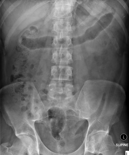
Analysis and Diagnosis
D – This is an Abdominal X-Ray taken on ….., of the following patient….. Is there a previous AXR to compare to?
R – Commenting first on the quality, it is not rotated, there is adequate field of view, the projection is anterior-posterior, the patient is lying supine and it is adequately exposed as I can see the vertebral bodies clearly
“On initial inspection, there appears to be signs of inflammation in the transverse colon, but I will proceed to go through it systematically.”
B – Looking at the film, the small bowel is not visible. There is loss of haustra markings in the transverse and descending colon. This looks like the “lead-pipe” sign which is suggestive of inflammation. There is also evidence of faecal loading in the ascending colon. The bowels are not dilated and no sign of obstruction.
O – Looking at the other organs, there is no basal lung consolidation. No hepatobiliary abnormality. The urinary system looks normal and no evidence of a psoas muscle abscess.
B – There are no fractures to the bones. There is a lateral curvature of the spine but the vertebrae appear normal. There is normal spacing of the sacroliliac and hip joints.
C– There are no other calcifications or foreign bodies.
In summary this AXR shows some inflammatory changes in the large bowel with loss of hautra markings and faecal loading in the ascending colon. It would be useful to see if there is previous to compare to in order to see if this is a new finding or chronic.
Diagnosis
Lead pipe colon and faecal loading in ascending colon – found in ulcerative colitis
Example 2
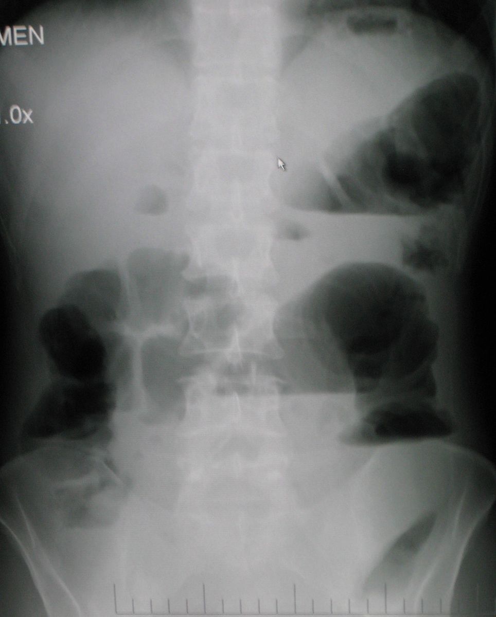
Diagnosis
Small Bowel Obstruction
Example 3
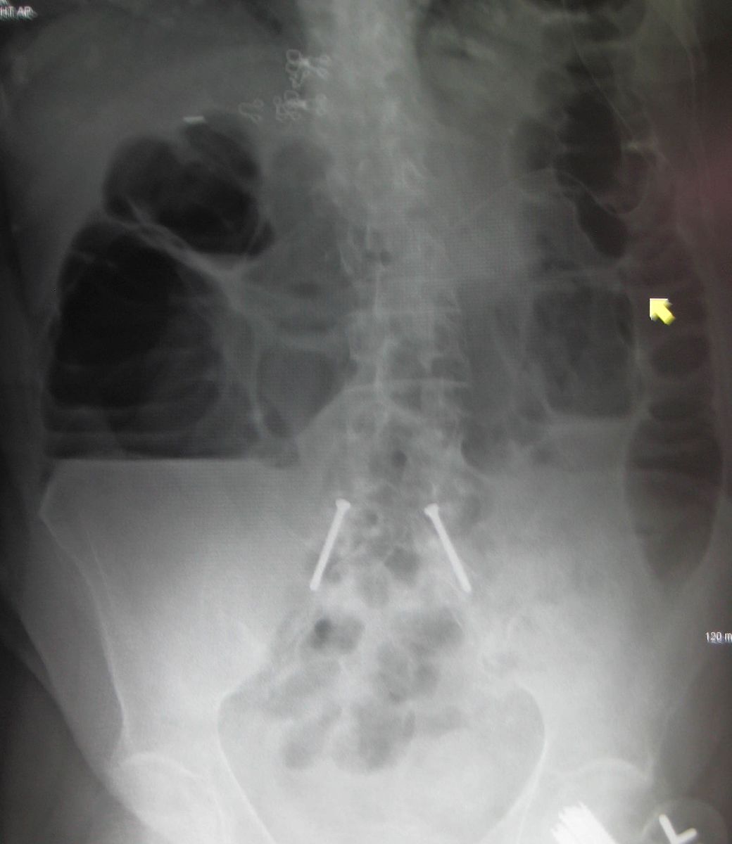
Diagnosis
Large Bowel Obstruction
Example 4
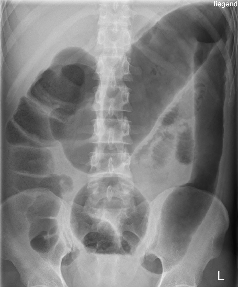
Diagnosis
Toxic Megacolon
Example 5
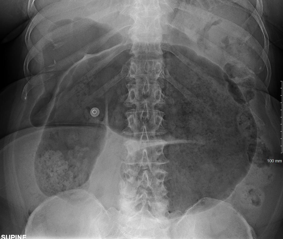
Diagnosis
Caecal Volvulus (“embryo sign”)
Check out the following pages to see more examples of common pathologies seen on abdominal X-Ray.
Sources
Image 2: James Heilman, MD, CC BY-SA 3.0
Image 3:James Heilman, MD, CC BY-SA 3.0
Image 4:Hellerhoff, CC BY-SA 3.0
Image 5: James Heilman, MD, CC BY-SA 4.0
Disclaimer

