Other Conditions
Check out these examples of abdominal X-Rays. Have a read of the first example where we go through how to present this plain film.
Practice with the remaining examples and click on the box to reveal the diagnosis.
Example 1
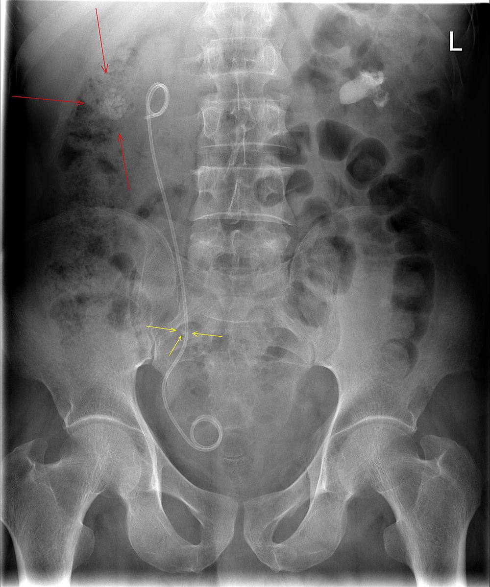
Take a look at the following example. Let us go through how we would systematically analyse this and the diagnosis.
Analysis and Diagnosis
D – This is an Abdominal X-Ray taken on ….., of the following patient….. Is there a previous AXR to compare to
R – Commenting first on the quality, it is not rotated, there is adequate field of view, the projection is anterior-posterior it is adequately exposed as I can see the vertebral bodies clearly
“On initial inspection, there is a ureteric stent in situ in the right ureter, but I will proceed to go through it systematically.”
B – Looking at the film, the small bowel is visible but it is not dilated. There is evidence of some faecal loading in the ascending colon. No evidence of bowel obstruction.
O – Looking at the other organs, there is no basal lung consolidation. No hepatobiliary abnormality. There is evidence of calcification in the right and left kidneys as well as a possible stone in the right ureter.
B – There are no fractures to the bones. There is a lateral curvature of the spine but the vertebrae appear normal. There is normal spacing of the sacroliliac and hip joints.
C– There are bilateral calcifications in the kidneys.
In summary this AXR shows some faecal loading in the ascending colon, bilateral calcifications in the kidneys and a right sided ureteric stent in situ.
Diagnosis
Right sided ureteric Stent
Example 2
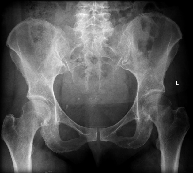
Take a look at the following example showing consolidation. Click on the box to reveal the diagnosis.
Diagnosis
Neck of femur fracture
Example 3
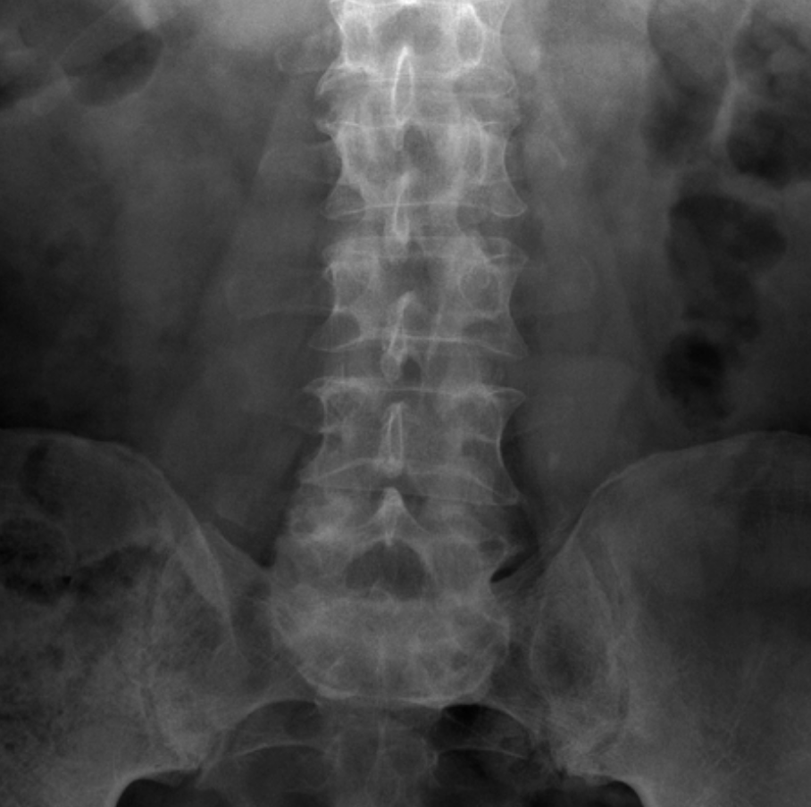
Diagnosis
Ureteric Stones
Example 4
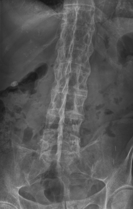
Diagnosis
Ankylosing Spondylitis (bamboo spine)
Example 5
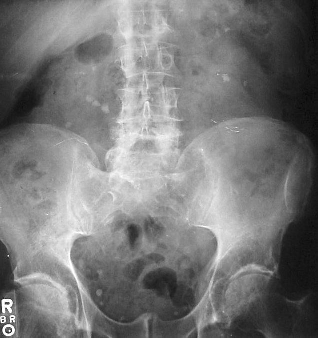
Diagnosis
Kidney Stones
Check out the following pages to see more examples of common pathologies seen on abdominal X-Ray.
Sources
Image 1: Lucien Monfils, CC BY-SA 3.0 <https://creativecommons.org/licenses/by-sa/3.0>, via Wikimedia Commons
Image 2: Gaillard, F., Bell, D. Neck of femur fracture. Reference article, Radiopaedia.org. (accessed on 11 Oct 2022) https://doi.org/10.53347/rID-1926
Image 3: Dr Graham Lloyd-Jones BA MBBS MRCP FRCR, AXR Tutorials < https://www.radiologymasterclass.co.uk/tutorials/abdo/abdomen_xray_calcium/calcium_ureteric_calcification#:~:text=Ureteric%20stones%20(calculi)%20are%20often,stones%20originate%20as%20renal%20stones.>
Image 4:Stevenfruitsmaak, CC BY-SA 3.0 <https://creativecommons.org/licenses/by-sa/3.0>, via Wikimedia Commons
Image 5: Bill Rhodes from Asheville, CC BY 2.0 <https://creativecommons.org/licenses/by/2.0>, via Wikimedia Commons
Disclaimer

