Foreign Objects
Foreign objects are important findings in chest X-rays so it is vital that you can distinguish these objects from normal structures .
Example 1
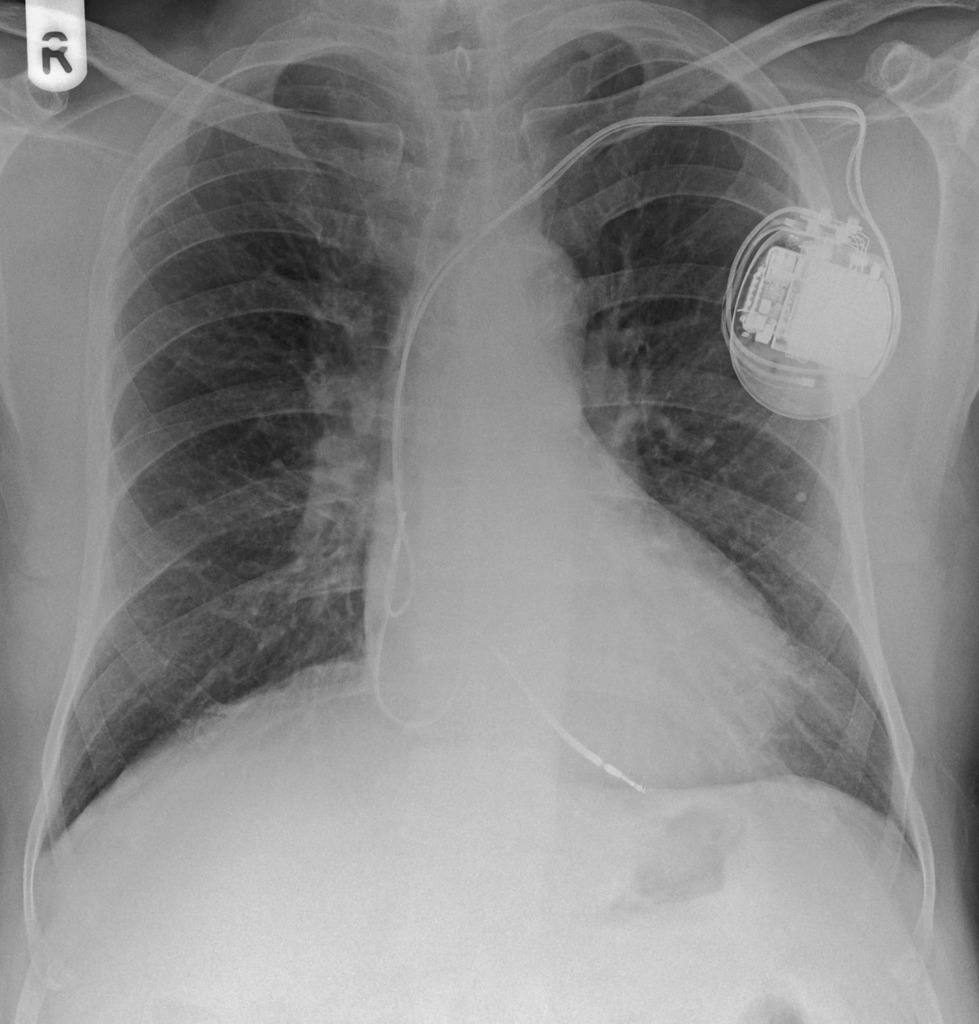
Take a look at the following example. Let us go through how we would systematically analyse this and the diagnosis.
Analysis and Diagnosis
D – This is a Chest X-Ray taken on ….., of the following patient….. Is there a previous CXR to compare to
R – Commenting first on the quality, it is not rotated, there is adequate inspiration, the projection is posterior-anterior
“On initial inspection, there appears to be a foreign object in the upper left chest, but I will proceed to go through it systematically.”
A – Starting with the airways, the trachea is not deviated, and the carina is visible.
B – The pleural markings go all the way to the costal margin so there is no evidence of a pneumothorax. Going through the lung zones, there is no opacification.
C – The heart is not enlarged and there is no loss of the heart border.
D – The hemidiaphragms are clearly visible and there is no blunting of the costophrenic angles. There is no free air under the diaphragm.
E – There is a pacemaker present, situated under the left clavicle. There are 2 wires which attach to the right atrium and the left ventricle.
There is no abnormality in the review areas, including the apices, behind the heart.
In summary the key finding is that there’s a pacemaker in situ
Diagnosis
Pacemaker
Example 2
Diagnosis
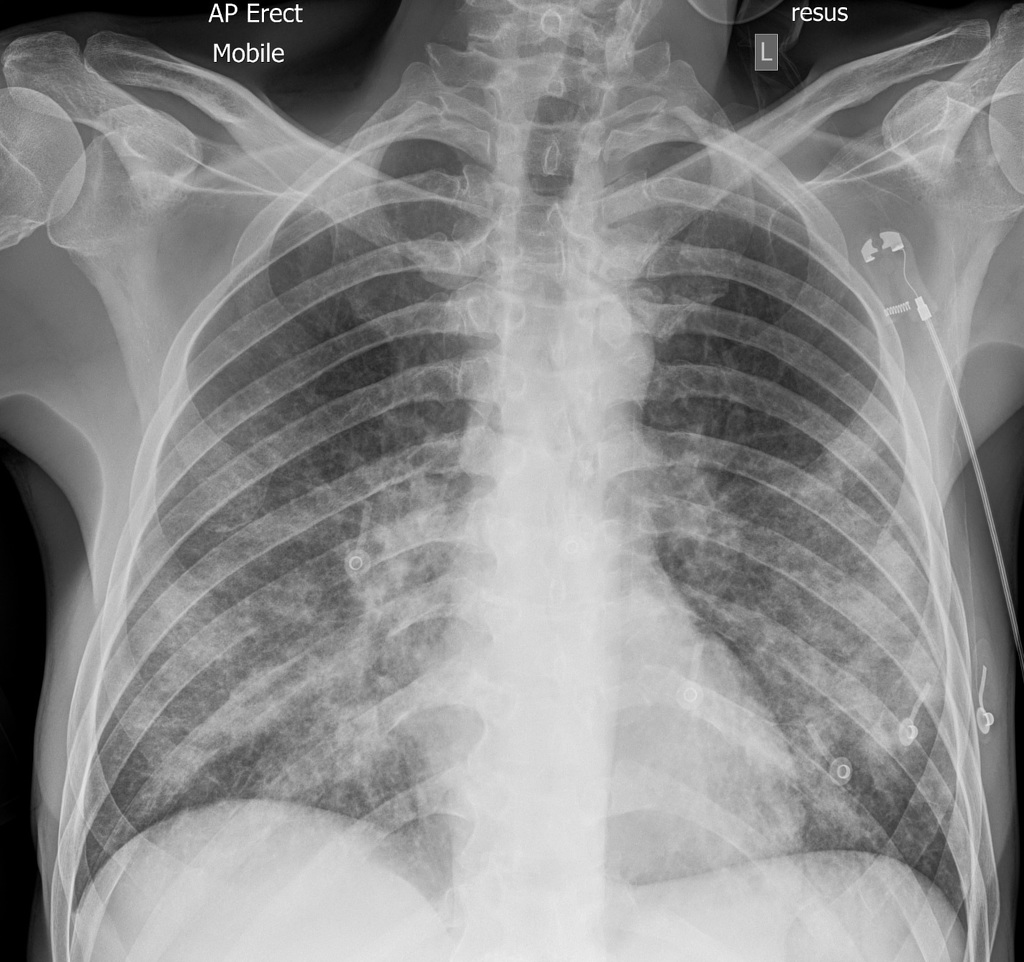
ECG
Example 3
Diagnosis
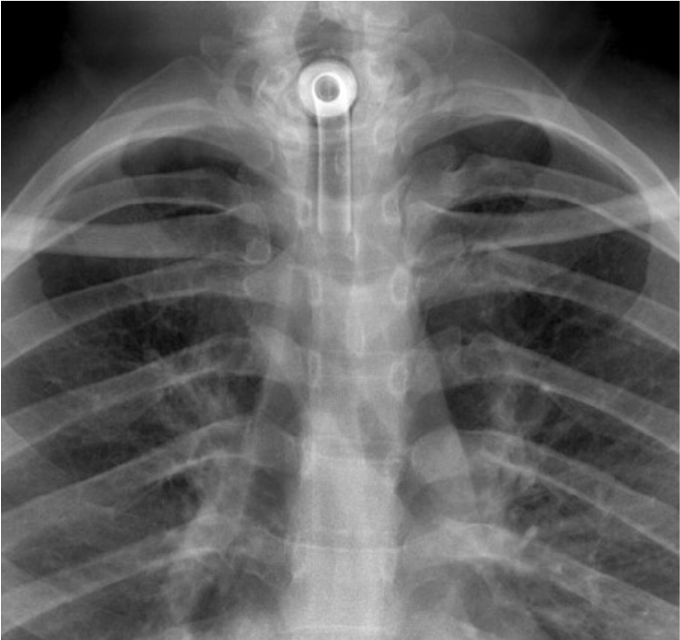
Endotracheal Tube
Example 4
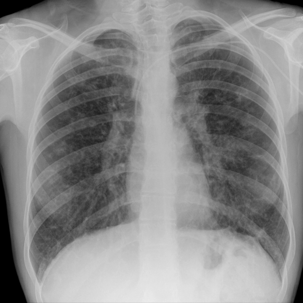
Diagnosis
PICC line
Example 5
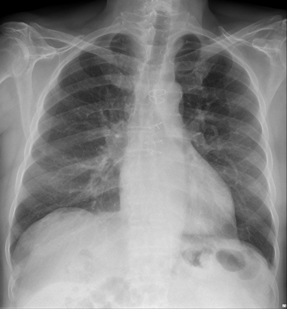
Diagnosis
Median Sternotomy Wires
Example 6
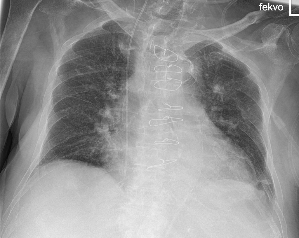
Diagnosis
Central Line and median sternotomy wires
Sources
Image 1:
http://www.wikiradiography.net/page/File:O2ZWd9HKVfas1Jyj3Ft1lw94915.jpeg#metadata
Image 2:
Bell, D. ECG chest leads. Case study, Radiopaedia.org. (accessed on 11 Oct 2022) https://doi.org/10.53347/rID-91649
Image 3:
Dr Graham Lloyd-Jones BA MBBS MRCP FRCR – Consultant Radiologist – Salisbury NHS Foundation Trust UK https://www.radiologymasterclass.co.uk/tutorials/chest/chest_tubes/chest_xray_et_tubes_anatomy
Image 4:
Jones, J. PICC line. Case study, Radiopaedia.org. (accessed on 11 Oct 2022) https://doi.org/10.53347/rID-6444
Image 5:
Knipe, H. Migrated sternotomy wire. Case study, Radiopaedia.org. (accessed on 11 Oct 2022) https://doi.org/10.53347/rID-55804
Image 6:
Botz, B. Deeply positioned central venous catheter. Case study, Radiopaedia.org. (accessed on 11 Oct 2022) https://doi.org/10.53347/rID-97221
Disclaimer

